
The serratus posterior inferior muscle, also known as the posterior serratus muscle,[citation needed] is a muscle of the human body.
Structure
The muscle is situated at the junction of the thoracic and lumbar regions.[1] It has an irregularly quadrilateral form, broader than the serratus posterior superior muscle, and separated from it by a wide interval.
It arises by a thin aponeurosis from the spinous processes of the lower two thoracic and upper two or three lumbar vertebrae.[1]
Passing obliquely upward and lateralward, it becomes fleshy, and divides into four flat digitations.[1] These are inserted into the inferior borders of the lower four ribs, a little beyond their angles.[1]
The thin aponeurosis of origin is intimately blended with the thoracolumbar fascia, and aponeurosis of the latissimus dorsi muscle.[citation needed]
Function
The serratus posterior inferior draws the lower ribs backward and downward to assist in rotation and extension of the trunk.[1] This movement of the ribs may also contribute to inhalation and forced expiration of air from the lungs.[2]
Additional images
-
 Position of the serratus posterior inferior (shown in red). Animation.
 -
 Close up. The muscle arises from the vertebrae T11 through L2 and inserted into lower border of the 9th through 12th ribs.
 -
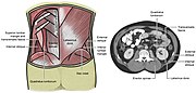 Lumbar triangle


See also
References
![]() This article incorporates text in the public domain from page 404 of the 20th edition of Gray's Anatomy (1918)
This article incorporates text in the public domain from page 404 of the 20th edition of Gray's Anatomy (1918)
- ^ a b c d e Jolley, C. J.; Moxham, J. (January 1, 2006), "RESPIRATORY MUSCLES, CHEST WALL, DIAPHRAGM, AND OTHER", in Laurent, Geoffrey J.; Shapiro, Steven D. (eds.), Encyclopedia of Respiratory Medicine, Oxford: Academic Press, pp. 632–643, ISBN 978-0-12-370879-3, retrieved January 17, 2021
- ^ Chaitow, Leon; DeLany, Judith (January 1, 2011), Chaitow, Leon; DeLany, Judith (eds.), "Chapter 10 - The lumbar spine", Clinical Application of Neuromuscular Techniques, Volume 2 (Second Edition), Oxford: Churchill Livingstone, pp. 211–297, ISBN 978-0-443-06815-7, retrieved January 17, 2021
External links
- Anatomy figure: 01:05-04 at Human Anatomy Online, SUNY Downstate Medical Center - "Intermediate layer of the extrinsic muscles of the back, deep muscles."
- Cross section image: pembody/body8a—Plastination Laboratory at the Medical University of Vienna

Recent Comments