
149.173.6.51 (talk) No edit summary |
Eric Kvaalen (talk | contribs) Started sections on disorders and uses. |
||
| Line 42: | Line 42: | ||
The fundus of the urinary bladder is the superior surface of the bladder. It is [[lymphatically]] drained by the [[external iliac lymph nodes]]. The peritoneum lies superior to the fundus. |
The fundus of the urinary bladder is the superior surface of the bladder. It is [[lymphatically]] drained by the [[external iliac lymph nodes]]. The peritoneum lies superior to the fundus. |
||
== |
==Disorders== |
||
Disorders of or related to the bladder include: |
|||
| ⚫ | |||
* [[Overactive bladder]], a condition which affects a large number of people. |
|||
| ⚫ | |||
| ⚫ | |||
* [[Bladder cancer]] |
* [[Bladder cancer]] |
||
* [[Bladder infection]] |
|||
* [[Bladder spasm]] |
* [[Bladder spasm]] |
||
* [[Bladder sphincter dyssynergia]], a condition in which the sufferer cannot coordinate relaxation of the urethra sphincter with the contraction of the bladder muscles |
* [[Bladder sphincter dyssynergia]], a condition in which the sufferer cannot coordinate relaxation of the urethra sphincter with the contraction of the bladder muscles |
||
* [[Bladder stone]]s |
|||
* [[Cystitis]] |
* [[Cystitis]] |
||
* [[Hematuria]], or presence of blood in the urine, is a reason to seek medical attention without delay, as it is a symptom of bladder cancer as well as bladder and [[kidney stone]]s. |
* [[Hematuria]], or presence of blood in the urine, is a reason to seek medical attention without delay, as it is a symptom of bladder cancer as well as bladder and [[kidney stone]]s. |
||
| ⚫ | |||
* [[Bladder exstrophy]] |
|||
==Uses== |
|||
Besides its normal use to the possessor, animal bladders (usually [[pig]] bladders) have been used to make balls (such as [[football (ball)|football]]s, and even a musical instrument, the [[bumbass]]. |
|||
| ⚫ | |||
| ⚫ | |||
* [[Bladder augmentation]] |
|||
* [[Neurogenic bladder]] |
* [[Neurogenic bladder]] |
||
* [[Ureterocele]] |
* [[Ureterocele]] |
||
| ⚫ | |||
* [[Urodynamics]] The study of the functional aspects of the detrusor muscle. |
* [[Urodynamics]] The study of the functional aspects of the detrusor muscle. |
||
* [[Uvula of urinary bladder]] |
* [[Uvula of urinary bladder]] |
||
* [[Vesicouretic reflux]] |
* [[Vesicouretic reflux]] |
||
* [[Urinary bladder disease]] |
* [[Urinary bladder disease]] |
||
==References== |
|||
| ⚫ | |||
==External links== |
==External links== |
||
Revision as of 08:32, 27 July 2008
In anatomy, the urinary bladder is a hollow, muscular, and distensible (or elastic) organ that sits on the pelvic floor in mammals. It is the organ that collects urine excreted by the kidneys prior to disposal by urination. Urine enters the bladder via the ureters and exits via the urethra.
In males, the bladder is superior to the prostate, and separated from the rectum by the rectovesical excavation.
In females, the bladder is separated from the uterus by the vesicouterine excavation.
Detrusor muscle
The detrusor muscle is a layer of the urinary bladder wall made of smooth muscle fibers arranged in spiral, longitudinal, and circular bundles. When the bladder is stretched, this signals the parasympathetic nervous system to contract the detrusor muscle. This encourages the bladder to expel urine through the urethra.
For the urine to exit the bladder, both the autonomically controlled internal sphincter and the voluntarily controlled external sphincter must be opened. Problems with these muscles can lead to incontinence. If the amount of urine reaches 100% of the urinary bladder volume, the voluntary sphincter becomes involuntary and the urine will be ejected instantly. The body cannot afford having the urinary bladder burst.
The urinary bladder usually holds 400–620 mL of urine, but it can hold twice this without rupturing if, for example, the outflow is obstructed.
The desire to urinate usually starts when the bladder reaches around 75% of its working volume. If the subject is distracted the desire can fade and return with more urgency as the bladder continues to fill.
Since the urinary bladder has a transitional epithelium, compared to the intestine mucosa, the urinary bladder does not produce mucus. [1]
Fundus
The fundus of the urinary bladder is the superior surface of the bladder. It is lymphatically drained by the external iliac lymph nodes. The peritoneum lies superior to the fundus.
Disorders
Disorders of or related to the bladder include:
- Overactive bladder, a condition which affects a large number of people.
- Bladder cancer
- Bladder infection
- Bladder spasm
- Bladder sphincter dyssynergia, a condition in which the sufferer cannot coordinate relaxation of the urethra sphincter with the contraction of the bladder muscles
- Bladder stones
- Cystitis
- Hematuria, or presence of blood in the urine, is a reason to seek medical attention without delay, as it is a symptom of bladder cancer as well as bladder and kidney stones.
- Urinary incontinence
- Bladder exstrophy
Uses
Besides its normal use to the possessor, animal bladders (usually pig bladders) have been used to make balls (such as footballs, and even a musical instrument, the bumbass.
See also
- Artificial bladder
- Bladder augmentation
- Neurogenic bladder
- Ureterocele
- Urodynamics The study of the functional aspects of the detrusor muscle.
- Uvula of urinary bladder
- Vesicouretic reflux
- Urinary bladder disease
References
External links
- Histology at KUMC epithel-epith09 "Urinary Bladder"
- Anatomy photo: Urinary/mammal/bladder/bladder1 - Comparative Organology at University of California, Davis - "Mammal, bladder (LM, Medium)"
- Template:IowaHistologyInteractive
- Anatomy photo:43:07-0100 at the SUNY Downstate Medical Center - "The Female Pelvis: The Urinary bladder"
- Anatomy photo:44:04-0103 at the SUNY Downstate Medical Center - "The Male Pelvis: The Urinary bladder"
Additional images
-
 Structure of the penis
 -
 Organs of the female reproductive system.
 -
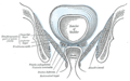 Coronal section of pelvis, showing arrangement of fasciæ. Viewed from behind.
 -

-
 The peritoneum of the male pelvis.
 -
 Median sagitta section of male pelvis.
 -
 Male pelvic organs seen from right side.
 -
 Median sagittal section of female pelvis.
 -
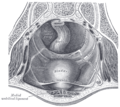
 The interior of bladder.
 -
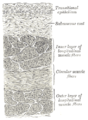 Vertical section of bladder wall.
 -
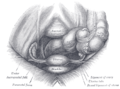
 Fundus of the bladder with the vesiculæ seminales.
 -
 Vertical section of bladder, penis, and urethra.
 -
 Female pelvis and its contents, seen from above and in front.
 -
 Topography of thoracic and abdominal viscera.
 -
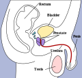 The bladder can be seen highlighted in yellow in the illustration.
 -
 Layers of the urinary bladder wall and cross section of the detrusor muscle.


Recent Comments